

 Flexural deformities of the forelimbs in horses can be either congenital (present at birth) or acquired (develops over time as the young horse grows, or secondary to severe injury).
Acquired flexural deformities of both the fetlock and coffin joints are a relatively common finding in Quarter Horses, along with many other breeds. Acquired deformity of the fetlock in the forelimb can be seen at different levels of severity, from very upright conformation to “knuckling over” (the fetlock joint will be positioned dorsal to or in front of the foot) at rest only (can lock the fetlock back into an extended position when walked) to “knuckling over” at all times (unable to extend the fetlock joint at all). The cause of acquired flexural deformities is thought to consist of multiple factors including growth rate, pain in the flexor tendons, amount of exercise (exacerbating pain in the flexor tendons), and trimming/shoeing practices.
The growth rate theory is related to the idea that the bones in the leg are growing in length at a faster rate than the body’s tendons and ligaments can catch up. In conjunction with that, as the tendons and ligaments attempt to stretch to keep up with bone growth, the horse begins to experience some pain in those tissues. This creates a repetitive cycle, as when the tendons are painful the horse does not want to bear full weight or extend the joints of the distal limb, further facilitating the flexed conformation. Alternatively, pain in the flexor tendons can be a primary causative factor rather than secondary.
In some cases, acquired flexural deformities of the fetlock can be seen in older horses (not young, growing horses) secondary to a chronic injury or painful condition in the affected limb. As the horse continues to not bear weight on the painful limb, tendon contracture takes place over time, shortening the musculotendinous unit in comparison to the bone and resulting in a joint region that is fixed in a flexed position.
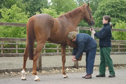 Diagnosis of flexural deformity of the fetlock joint is principally made based on clinical exam and visualization of the deformity. There is a wide range of treatment approaches, from more conservative methods to surgical procedures. Whether surgical treatment is considered or not, nutritional factors should always be considered in the treatment plan, as controlling nutrition can aid in controlling growth rate. For older, weanling foals and yearlings, eliminating or minimizing concentrate (grain) from the diet will be critical. The horse should be restricted to good quality grass hay; the goal should be to restrict the diet to the point where the horse is not overly thin, but that the ribs are slightly visible. For younger foals, early weaning is often helpful as a means to decrease the plane of nutrition. Exercise restriction is often beneficial as well, based on the concept that pain in the flexor tendons is a contributing factor. Along with exercise restrictions, treatment of pain with non-steroidal anti-inflammatory drugs (Phenylbutazone) will be beneficial to treat or control any pain from increased tension on the flexor tendons. Because flexural deformity of the fetlock is often attributed to a functional shortening of the superficial digital flexor specifically, corrective shoeing by applying a wedge may be beneficial. The wedge will reduce some strain on the deep digital flexor tendon while increasing strain on the superficial digital flexor tendon (increasing stretch on the tendon), however control or treatment of a secondary pain component must remain a part of the treatment plan. Splint application may be beneficial as well; the splint is applied down the back or palmar side of the entire forelimb, with effort fixed on holding the fetlock joint in an extended position. Again, control of pain in the flexor tendons must be considered.
For more severe or chronic cases, or those that have failed to respond to conservative approaches, surgical management should be considered. Typically the superior check ligament, or accessory ligament of the superficial digital flexor tendon, is surgically transected; this ligament originates from the caudal aspect of the distal radius, and joins the superficial digital flexor tendon/muscle, so rapid growth of the radius is thought to result in a shortening of this musculotendinous unit. In some cases (based on which of the flexor tendons is more taut when the fetlock is extended), the inferior check ligament (or accessory ligament of the deep digital flexor tendon, traditionally cut for flexural deformities of the coffin joint) is transected instead. In more severe cases, surgical transection of both the superior and inferior check ligaments is performed. Post-operative treatments include full limb splinting and/or application of a wedge shoe as described above. Two surgical approaches have been developed, an open approach and a minimally invasive endoscopic approach. The minimally invasive endoscopic approach, developed in an effort to minimize scarring, white hairs, and other incisional complications associated with the open approach (hemorrhage, seroma development, swelling), involves making two small incisions into the carpal sheath, and cutting the ligament under endoscopic guidance. The arthroscope/endoscope is placed into one incision, and an instrument for cutting placed into the second. Cosmetic outcome following the minimally invasive approach is excellent (particularly if wedge shoes only are used post surgery, as splinting tends to result in white hairs over time), and clinical results are comparable to those seen with the traditional open approach. As described above, dietary restrictions, exercise restriction, splinting and/or wedge shoes, and pain control are still critical components to the post-operative management of these cases.
Megan Williams graduated from Kansas State University and went on to an internship at Ocala Equine Hospital. She completed her surgical residency at Michigan State University. Dr. Williams is a surgeon at Saginaw Valley Equine Clinic and a lameness diagnostician with GameTime Sports Medicine at major AQHA events around the country. You can email her at megan@saginawvalleyequine.com or visit her at www.saginawvalleyequine.com. You can also write to her in care of InStride Edition.
You must be logged in to post a comment Login